报 告 人:Angela Ruohao WU, 香港科技大学教授
报告题目:Investigating human tissue heterogeneity using quantitative single-cell transcriptomics
报 告 人:黄岩宜, 北京大学教授,国家杰出青年基金获得者
报告题目:Highly accurate and precise single-cell sequencing facilitated by microfluidics
邀请人: 刘笔锋 教授
报告时间:2016年4月12日星期二上午9:30
报告地点:武汉光电国家实验室 A302
Abstract(Prof. Angela Ruohao WU):
The field of single-cell whole transcriptome analysis is growing rapidly to achieve profiling of rare or heterogeneous populations of cells. We compared the performance of various commercially available single-cell RNA amplification methods in both microliter and nanoliter volumes. We benchmarked each method to conventional RNA-seq of the same sample using bulk total RNA, as well as to multiplexed qPCR. In doing so, we were able to systematically evaluate the sensitivity, precision, and accuracy of various approaches to single-cell RNA-seq. Our results show that it is possible to use single-cell RNA-seq to perform quantitative transcriptome measurements of single cells, that it is possible to obtain useful gene expression measurements with a relatively small number of sequencing reads, and that when such measurements are performed on large numbers of cells, one can recapitulate both the bulk transcriptome complexity as well as the distributions of gene expression levels found by single-cell qPCR. We subsequently used microfluidic-based RNA-seq to profile single cells of the developing mouse lung, and adult human colon respectively to dissect their tissue heterogeneities, discover novel cell types, as well as investigate biological mechanisms.
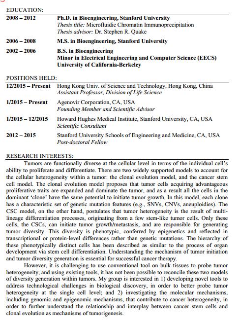
About Angela Ruohao WU, Ph.D

Abstract (黄岩宜教授):
Phenotype classification of single cells reveals biological variation that is masked in ensemble measurement. This heterogeneity is found in genomic variation and gene expression as well as in cell morphology. Many techniques are available to probe phenotypic heterogeneity at the single cell level, for example single-cell genomic sequencing, RNA sequencing, and quantitative imaging, but it is difficult to perform multiple assays on the same single cell.
On one hand, to better assessing the genomic variations between single cells, we developed a microfluidic-based emulsion whole genome amplification (eWGA) method to overcome the technical limitations existing in prevailing single-cell WGA approaches. We divide single-cell genomic DNA into a large number of picoliter aqueous droplets in oil. This easy-to-operate approach enables simultaneous detection of CNVs and SNVs in an individual human cell, exhibiting significantly improved amplification evenness and accuracy.
On the other hand, in order to directly track correlation between gene expression and morphology of each single cell, we developed a microfluidic platform for quantitative label-free imaging and immediate RNA sequencing (RNA-Seq) of single cells. With this device we actively screen and trap cells for analysis with coherent Raman microscopic imaging. The cells are then processed in parallel pipelines for lysis, and preparation of cDNA for high-throughput transcriptome sequencing. Coherent Raman microscopy offers three-dimensional imaging with chemical specificity for quantitative analysis of protein and lipid distribution in single cells. Meanwhile, the microfluidic platform facilitates single-cell manipulation, minimizes contamination, and furthermore, provides improved RNA-Seq detection sensitivity and measurement precision, which is necessary for differentiating biological variability from technical noise.
Biography:
Yanyi Huang received his Bs (Chemistry) and ScD (Inorganic Chemistry) from Peking University in 1997 and 2002, respectively. He did his postdoc works with Amnon Yariv at Caltech (Applied Physics, 2002-2005) and with Stephen Quake at Stanford (Bioengineering, 2005-2006). He joined Peking University faculty in 2006. He is Professor of Materials Science and Engineering, Principal Investigator of Biodynamic Optical Imaging Center, Principal Investigator of Peking-Tsinghua Center for Life Sciences.